|
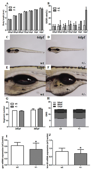
Developmental parameters and mRNA expression of developmental genes in fushi tarazu factor 1b (ff1b) knockout zebrafish by CRISPR/Cas9 technology. (A-D) Developmental parameters of F3 zebrafish embryos; (A) body lengths; (B) intrauterine growth retardation (IUGR) rates; (C) the whole body of F3 wild-type (WT) embryos at 6 days post-fertilization (dpf); (D) the whole body of F3 mutant-type (MT) embryos at 6 dpf; (E) the yolk and swim bladder of F3 WT embryos at 6 dpf; (F) the yolk and swim bladder of F3 MT embryos at 6 dpf. White arrow: swim bladder; black arrow: yolk. (G) Segment number; (H) general morphology score (GMS). Mean ± SD, *P<0.05, two-sided t-test, n = 3 F2 zebrafish. Real-time reverse-transcription PCR (RT-PCR) was used to detect the mRNA expression of developmental genes in F3 embryos. (I) goosecoid (gsc) expression in F3 embryos at 12 hours post-fertilization (hpf); (J) krox20 expression in F3 embryos at 12 hpf. Mean ± SD, *P<0.05, two-sided t-test, n = 3 embryos.
|

