
Fig. 6
- ID
- ZDB-FIG-180222-23
- Publication
- Akerberg et al., 2017 - Histone demethylases Kdm6ba and Kdm6bb redundantly promote cardiomyocyte proliferation during zebrafish heart ventricle maturation
- Other Figures
- All Figure Page
- Back to All Figure Page
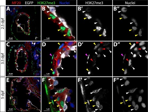
Bulk cellular H3K27me3 levels transiently decrease in trabeculating cardiomyocytes. (A-F) Confocal microscopy immunofluorescence images of sagittal sections through the heart of 2.5, 3.5, and 5 dpf Tg(kdrl:EGFP) embryos stained with anti-myosin heavy chain (red, MF20, myocardium), anti-EGFP (white, endocardium), and anti-H3K27me3 (green) antibodies. Hoechst-stained nuclei are blue. Zoomed images of the yellow boxed areas (in A, C, and E) are shown in (B, D, and F) with adjacent single channel H3K27me3 and nuclei staining to highlight differences in bulk H3K27me3 levels. Magenta and white arrowheads indicate nuclei of epicardial and endocardial cells, respectively. Yellow and red arrowheads mark H3K27me3-high and H3K27me3-low cardiomyocyte nuclei, respectively. 20 μM and 5 μM (zoom panels) scale bars are shown. |
Reprinted from Developmental Biology, 426(1), Akerberg, A.A., Henner, A., Stewart, S., Stankunas, K., Histone demethylases Kdm6ba and Kdm6bb redundantly promote cardiomyocyte proliferation during zebrafish heart ventricle maturation, 84-96, Copyright (2017) with permission from Elsevier. Full text @ Dev. Biol.