
Fig. 5
- ID
- ZDB-FIG-180103-42
- Publication
- Yan et al., 2017 - RETRACTED: Chemical inhibition reveals differential requirements of signaling pathways in krasV12- and Myc-induced liver tumors in transgenic zebrafish.
- Other Figures
- All Figure Page
- Back to All Figure Page
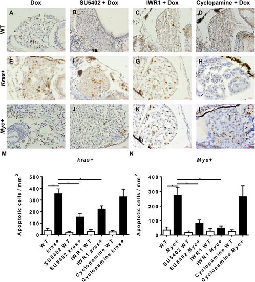
Cell apoptosis analysis of krasV12- and Myc-induced carcinogenesis. 7 dpf WT, kras+ or Myc+ larvae were treated with 10 μM SU5402, 10 μM IWR1 or 10 μM cyclopamine in the presence of 10 μg/ml Dox. Apoptosis was analyzed by immunohistochemical staining with digoxigenin-conjugated nucleaotide and incubated with anti-digoxigenin secondary antibody. (A–D) Representative liver images of 7 dpf WT larvae. (E–H) Representative liver images of 7 dpf kras+ larvae. (I–L) Representative liver images of 7 dpf Myc+ larvae. (M) Statistical analysis of numbers of apoptotic cells in the liver of kras + larvae. (N) Statistical analysis of numbers of apoptotic cells in the liver for Myc+ larvae. N = 20 from each groups; statistical significance: *p < 0.05, Scale bar = 20 μm. |