
Fig. 4
- ID
- ZDB-FIG-171006-21
- Publication
- Bostaille et al., 2016 - Molecular insights into Adgra2/Gpr124 and Reck intracellular trafficking
- Other Figures
- All Figure Page
- Back to All Figure Page
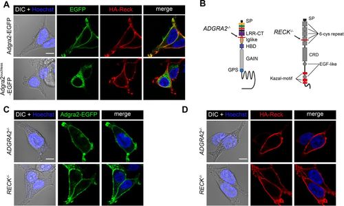
Independent trafficking of Reck and Adgra2 to the plasma membrane. (A) Single-plane confocal images of non-permeabilized HEK293T cells 48 h after transfection with HA-reck and adgra2-EGFP variants, as indicated. (B) Schematic representation of the genetic lesions of ADGRA2−/− and RECK−/− cells. The position of the frame-shift mutation is indicated by the red line. See Fig. 1A for schematic labels. (C,D) Single-plane confocal images of non-permeabilized ADGRA2−/− and RECK−/− HEK293T cells 48 h after transfection with adgra2-EGFP (C) and HA-reck (D) constructs. In all panels, EGFP is detected by direct fluorescence and the HA-Reck fusion by anti-HA indirect immunofluorescence. Cells were additionally transfected with a Wnt7a (mouse gene) expression construct. Nuclei were counterstained with Hoechst. Scale bars: 10 μm. |