
Fig. S6
- ID
- ZDB-FIG-170908-20
- Publication
- Breau et al., 2017 - Extrinsic mechanical forces mediate retrograde axon extension in a developing neuronal circuit
- Other Figures
- All Figure Page
- Back to All Figure Page
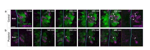
Morphologies of placodal cells expressing low levels of GFP. (a,b) Two time lapse sequences showing instances of central (a) and posterior (b) low ngn1:gfp+ cells undergoing lateral movements. Cells of interest are indicated with pink and yellow dots. The brain surface is indicated by a blue line. While moving laterally, the cells (labelled with mosaic mbCherry) extend a long cytoplasmic process contacting the brain surface (white arrowheads), as shown for high GFP-expressing cells in Figure 2. On the up right panel, note that all the protrusions of mbCherry-labelled placode cells meet to form a bundle on the brain surface {white dotted line). Scale bar: 25 µm . |