
Fig. S7
- ID
- ZDB-FIG-170908-21
- Publication
- Breau et al., 2017 - Extrinsic mechanical forces mediate retrograde axon extension in a developing neuronal circuit
- Other Figures
- All Figure Page
- Back to All Figure Page
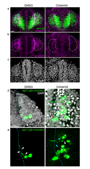
Additional results for colcemid treatment experiments. (a,b) ngn1:gfp embryos treated with colcemid or DMSO, and stained for acetylated tubulin at 24s. (a) shows the merge picture, and (b) the acetylated tubulin staining only. The acetylated tubulin appears affected and disorganised in the head of colcemid-treated embryos, and is strongly reduced in OPs (surrounded by white dotted lines). (c) XY sections of DAPI staining of the embryos shown in (a) and (b). Note the numerous nuclei blocked in mitosis in colcemid-treated embryos. These results confirm that colcemid treatment affects microtubule dynamics and metabolism. (d,e) Wild type embryos were injected with ngn1:gfp DNA to obtain a mosaic labelling of OP neurons, treated with colcemid or DMSO and stained for DAPI at 24s. In DMSO controls, mosaic labelling allows to visualise axonal protrusions connecting cell bodies to the brain surface (white arrows). In colcemid-treated embryos, some GFP+ cells possess a normal axonal protrusion (white arrow), but a significant proportion of GFP+ cells show shorter axons that do not contact the brain (yellow asterisks indicate the cell bodies of such cells, and blue arrows their short protrusions) or no axonal protrusion at all (white asterisks indicate their somata). Scale bars: 25 µm. |