
Fig. 6
- ID
- ZDB-FIG-170823-19
- Publication
- Yoo et al., 2016 - Plexins function in epithelial repair in both Drosophila and zebrafish
- Other Figures
- All Figure Page
- Back to All Figure Page
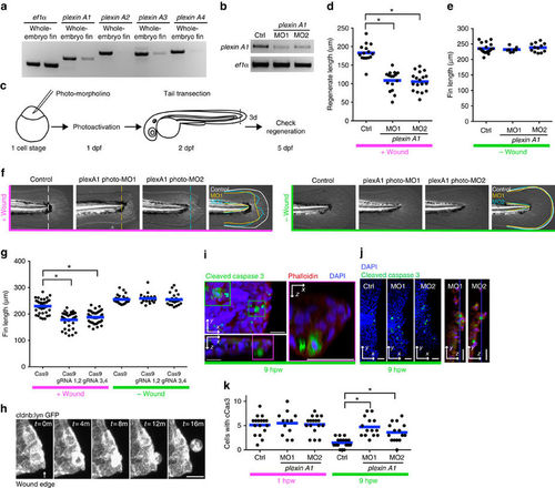
Plexin A1 regulates wound repair in zebrafish fins. (a) RT–PCR of plexins. plexin A1 and A3 are relatively enriched in tail fins at 2 d.p.f. (b) RT–PCR of plexin A1 following knockdown. Two independent splice morpholinos were used. (c) Diagram of photo-morpholino injection, photoactivation and tail transection. (d) Regenerated fin length at 3 days post wounding (d.p.w.), i.e., 5 d.p.f. *P<0.05; one-way ANOVA with Dunnett’s post-test. (e) Fin length at 5 d.p.f. in the absence of wounding. (f) Images of 5 d.p.f.-larval tail fins with/without wounding. Note that wounding affects both the length and width of the wounded tail fins in plexin A1 morphants. (g) CRISPR-mediated plexin A1 inhibition impairs fin regeneration at 3 d.p.w. (5 d.p.f.) but not the fin length without wounding. *P<0.05; one-way ANOVA with Dunnett’s post-test. (h) Epithelial cell extrusion around a wound of Tg(cldnb:lynGFP). On average, 2.75 (n=4, s.e.m.=0.48) cells were observed to be extruded for 5 h imaging, but note that all the extrusion events were not captured because imaging was done only in a part of the tail fin. (i) An extruded cell is undergoing apoptosis. (j) Representative images of transected tail fins at 9 h.p.w. (k) Knockdown of Plexin A1 increases apoptotic cells at wounds at 9 h.p.w. but not 1 h.p.w. *P<0.05; one-way ANOVA with Dunnett’s post-test. |
| Genes: | |
|---|---|
| Fish: | |
| Anatomical Terms: | |
| Stage: | Long-pec |
| Fish: | |
|---|---|
| Condition: | |
| Knockdown Reagents: | |
| Observed In: | |
| Stage: | Day 5 |