
Fig. 4
- ID
- ZDB-FIG-170413-17
- Publication
- Mandal et al., 2017 - Wnt signaling balances specification of the cardiac and pharyngeal muscle fields
- Other Figures
- All Figure Page
- Back to All Figure Page
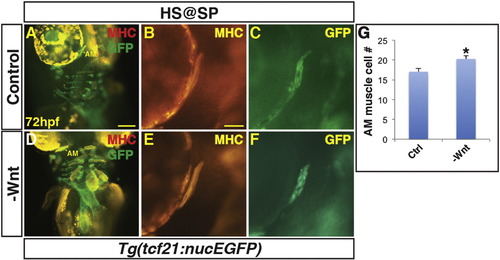
Decreased Wnt signaling causes an increase in 1st arch PM cells. (A, B) Tg(tcf21:nucEGFP) control sibling embryos and embryos with decreased Wnt signaling at the SP stage. Decreased Wnt signaling causes enlargement of the 1st arch AM muscle (boxes). Sarcomeric myosin (MHC; red). NucGFP (green). (B, E) MHC alone of AM muscles. (C, F) NucEGFP alone of AM. (G) Graph depicting quantification of nuclei in the AM muscles. Decreasing Wnt signaling at the SP stage produces a modest, but significant increase in AM muscle nuclei. Control embryos n = 15, Tg(hsp70l:dkk1-GFP) embryos n = 7. Asterisks in all graphs indicate p < 0.05. Error bars in all graphs indicate S.E.M. Scale bar in A indicates 100 μm. Scale bar in B indicates 50 μm. |
Reprinted from Mechanisms of Development, 143, Mandal, A., Holowiecki, A., Song, Y.C., Waxman, J.S., Wnt signaling balances specification of the cardiac and pharyngeal muscle fields, 32-41, Copyright (2017) with permission from Elsevier. Full text @ Mech. Dev.