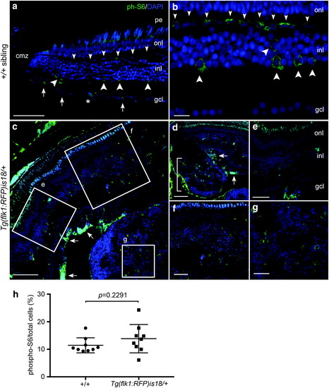
Activated mTOR signaling marker phosphorylated-S6 in adult zebrafish wild-type retina, Tg(flk1:RFP)is18/+ dysplastic retina, and retinal tumor. (a, b) phospho-S6 (green) is detected in putative horizontal cells (small arrowheads), amacrine and/or Müller glia (large arrowheads), and retinal ganglion cells and/or displaced amacrine cells (small arrows) in the mature region of the retina. Nuclei are labeled with 4′,6-diamidino-2-phenylindole. Asterisk marks a blood vessel. (c–g) In Tg(flk1:RFP)is18/+ dysplastic retina phospho-S6 labeling is present in a subset of cells throughout regions of the retina with increased cell number and disorganized retinal layers (c–f). Intense labeling of phospho-S6 was present in the ganglion cell and nerve fiber layers (c, d, arrows). Large lesions contained phospho-S6 positive cells distributed throughout the tumor mass (d [bracket], g). (h) Percentage of phospho-S6 cells/total cells in wild-type retina and Tg(flk1:RFP)is18/+ tumor. +/+ 11.4% ± 0.9 vs. Tg(flk1:RFP)is18/+ 13.9% ± 1.7, p = 0.2291. n = 9 sections, three sections each from three individuals of each genotype. gcl, ganglion cell layer; inl, inner nuclear layer; onl, outer nuclear layer; pe, pigmented epithelium. Scale bars, (a, g, f) 50 μm; (b, c) 20 μm; (d, e) 100 μm; (h) 25 μm.
|
