
Fig. 1
- ID
- ZDB-FIG-170317-1
- Publication
- Schultz et al., 2017 - Vascular Endothelial Growth Factor A and Leptin Expression Associated with Ectopic Proliferation and Retinal Dysplasia in Zebrafish Optic Pathway Tumors
- Other Figures
- All Figure Page
- Back to All Figure Page
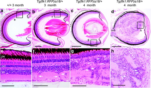
Detection of aberrant cell migration and ectopic proliferation in young adult Tg(flk1:RFP)is18 dysplastic retina. (a, e) Histological staining of retina section from 3-month-old adult wild-type zebrafish showing nuclear layers and organized laminar structure. (b, f) Representative image of section through retina of 3-month-old Tg(flk1:RFP)is18 adult reveals aberrant migration of cells across the inner nuclear layer (arrows, f) (n = 9). (c–h) Representative images of retinal sections from 4-month-old Tg(flk1:RFP)is18 adults (n = 15). (c, g) Retina from 4-month-old Tg(flk1:RFP)is18 adult with dysplasia reveals disruption of inner, outer, and ganglion cell layers with numerous mitotic figures (arrows, g). (d, h) Four-month-old Tg(flk1:RFP)is18 adult with advanced retinal tumor filling the vitreous space. Tumor tissue is composed of fibrous material interspersed with numerous mitotic figures, cells showing heterogeneous nuclear morphology and forming occasional rosettes (arrows, h), and blood vessels (arrowheads, h). pe, pigmented epithelium; onl, outer nuclear layer; inl, inner nuclear layer; gcl, ganglion cell layer. Scale bars (a, b) 200 μm; (c, d) 500 μm; (e–h) 50 μm. |
| Fish: | |
|---|---|
| Observed In: | |
| Stage: | Adult |