
Fig. 7
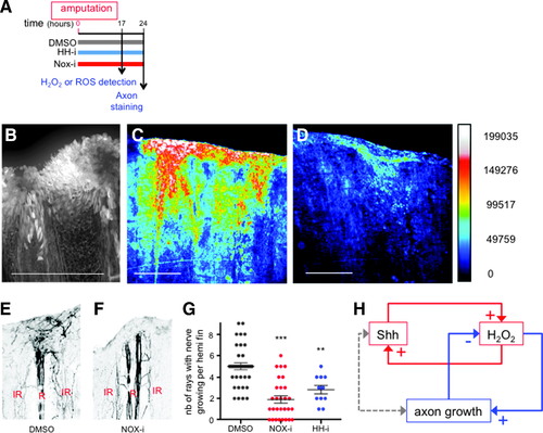
H2O2 stimulates axon growth in a positive feedback loop. (A) Scheme of the experiments. (B) ROS detection with DCFDA fluorescent probe. (C, D) Detection of H2O2 in ubi:HyPer fish. Fish were incubated in water (C) or Nox-i (D) and fluorescence of HyPer analyzed on adult anesthetized fish. [H2O2] is inferred from the YFP500/YFP420 excitation ratio of HyPer. Pseudocolor calibration bars: HyPer ratio (YFP500/YFP420). (E, F) Growing nerves were detected in the regenerating fin at 24 hpa by immunodetection of acetylated tubulin in fish incubated in vehicle (E), Nox-i (F), or HH-i (not shown). The quantification presented in (G) corresponds to the number of rays with growing nerves per half fin (maximum 9). n Values: dimethyl sulfoxide = 36; NOX-i = 29; HH-i = 11. (H) This article reveals the interaction between two feedback loops in the regulation of axon growth. Error bars represent the SEM (**p < 0.01; ***p < 0.001). Scale bars = 200 μM. YFP, yellow fluorescent protein. |