
Fig. 3
- ID
- ZDB-FIG-160906-4
- Publication
- Pipalia et al., 2016 - Cellular dynamics of regeneration reveals role of two distinct Pax7 stem cell populations in larval zebrafish muscle repair
- Other Figures
- All Figure Page
- Back to All Figure Page
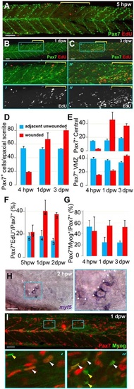
Rapid recovery of Pax7-expressing cells in wounded somites through proliferation and relocation enhances differentiation in central myotome. Wild-type zebrafish larvae wounded at 3dpf in epaxial somites 16-18 (yellow brackets) were analysed at the indicated times post-wounding by confocal immunodetection of Pax7 with EdU (A-F) or Myogenin (G,I) or in situ mRNA hybridisation for myf5 (H), shown in lateral view, anterior to left, dorsal to top. Blue boxes are magnified. (A-C) Diminished numbers of Pax7+ cells after wounding (A) are rapidly replaced (B) and show increased proliferation (B,C). (D-G). Pax7+ cells were counted in 2-4 wounded and 2-4 adjacent unwounded somite regions per larva and averaged to yield a value for each animal. VMZ, vertical myoseptum. Mean±s.e.m values from four larvae (D,E,G) or the number indicated (F). Statistical analysis is shown in Fig. S3A. (H) myf5 mRNA adjacent to a hypaxial wound (outlined by dots). Note the lack of myf5 mRNA in unwounded somites at this stage. (I) Pax7+Myog+ nuclei (white arrowheads) generally have lower Myog signal than Pax7-Myog+ cells (yellow arrowheads). Scale bars: 50µm. |
| Gene: | |
|---|---|
| Antibodies: | |
| Fish: | |
| Condition: | |
| Anatomical Terms: | |
| Stage Range: | Protruding-mouth to Day 6 |