
Fig. 4
- ID
- ZDB-FIG-160906-5
- Publication
- Pipalia et al., 2016 - Cellular dynamics of regeneration reveals role of two distinct Pax7 stem cell populations in larval zebrafish muscle repair
- Other Figures
- All Figure Page
- Back to All Figure Page
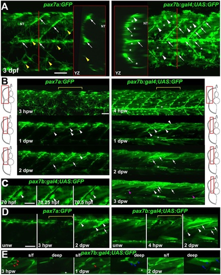
pax7a- and pax7b-reporter transgenes express distinctly during wound repair. (A) Confocal maximum intensity projection of stacks of whole somites in lateral view, showing the distribution of GFP+ cells at 3dpf in unwounded pax7a:GFP and pax7b:gal4;UAS:GFP fish. Note xanthophores (yellow arrowheads), dermomyotomal cells (white arrowheads), VMZ and HZM cells (white arrows), and labelled fibres (asterisks). Transverse YZ slices were taken at the red line. NT, neural tube. (B) Live confocal time lapse imaging of short stacks taken from the volumes indicated in the adjacent transverse schematics, shown in lateral view, anterior to left, dorsal up. Note loss of signal at 3-4hpw in regions wounded at 3dpf (yellow brackets), and recovery of GFP+ cells (arrowheads) and muscle fibres (arrows) over the ensuing days. Asterisk indicates persistence of a deep fast fibre marked by pax7b-reporter prior to wounding. Unwounded somites also accumulate small numbers of marked mononucleate cells (magenta arrowheads). (C) Time series confocal slices showing pax7b-reporter+ cell division (arrowheads) prior to wounding. (D) Magnified confocal slices showing wounding (yellow brackets) and repair. Note the stronger fibre labelling (arrows) with pax7b-reporter than with pax7a:GFP, relative to mononucleate cells (arrowheads). (E) Time series confocal slices showing superficial (s/f, left panels) and deep (right panels) pax7b-reporter+ cell appearance in the wound region followed by fusion. Disappearance of small bright GFP+ cells amongst the superficial fibres (red arrowheads) correlated with appearance of small bright GFP+ cells in deep regions (blue arrowheads, centre). Loss of some small deep cells then correlated with appearance of weakly GFP-labelled fibres (blue arrows; rightmost panel). Asterisks indicate a deep fast fibre marked by pax7b-reporter prior to wounding. Scale bars: 50µm. |
| Genes: | |
|---|---|
| Fish: | |
| Condition: | |
| Anatomical Terms: | |
| Stage Range: | Pec-fin to Day 6 |