|
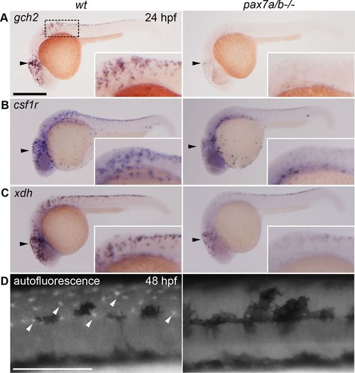
The pax7a/pax7b double-mutant embryos have a reduced xanthoblast pool and completely lack differentiated xanthophores. Whole-mount in situ hybridization on wt siblings and pax7a/pax7b double-mutant zebrafish embryo at 24 hpf showing the expression of (A) gch2 (n = 13 for siblings and n = 7 for pax7a/pax7b double mutants), (B) csf1r (n = 5 and 3), and (C) xdh (n = 3 and 5). (D) Pteridine autofluorescence in wt and pax7a/pax7b double-mutant zebrafish embryo at 48 hpf. Box indicates area of enlargement visualized in insets. Black arrowheads indicate head region where changes in expression can be detected; white arrowheads indicate a subset of autofluorescing xanthophores. Scale bar, 200 μm.
|