
Fig. 3
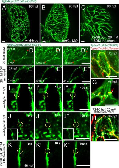
Cardiac contraction is required for Cdh2-EGFP clustering. Spinning disk confocal images of TgBAC(cdh2:cdh2-EGFP) hearts. Localization of Cdh2-EGFP at 96 hpf in wild-type (A) and stopped hearts using tnnt2a morpholino (B) or 20 mM BDM (C). (D-D′′ and E-E′′) Single plane images at different points. (D-D′′) Cdh2-EGFP in punctae became evenly distributed upon treatment with BDM. (E-E′′) Removal of BDM resulted in the reclustering of Cdh2-EGFP into a punctate pattern. (F-H) Spinning disk confocal images of Tg(myl7:LIFEACT-GFP);Tg(myl7:cdh2-tdTomato) hearts focusing on the basal side of compact layer cardiomyocytes. Red arrows point to colocalization of actin bundle and Cdh2 punctae. On BDM treatment, the association between Cdh2-GFP punctae and actin bundles appears disrupted. (I-K) Dynamics of Cdh2-EGFP localization revealed by fluorescence recovery after photobleaching. (I′′) Recovery of Cdh2-EGFP signal in a 52 hpf heart after photobleaching. (J′′) After photobleaching of Cdh2-EGFP puncta at 96 hpf, it was not able to reform even though a diffuse Cdh2-EGFP signal is observed in the bleached region; zoomed images from bleached region. (K′′) Recovery of Cdh2-EGFP signal in a nonbeating heart at 96 hpf after photobleaching. (Scale bars, 10 µm in A-C, D′′-E′′, and F-H; 5 µm in I′′-K′′.) |