
Fig. 2
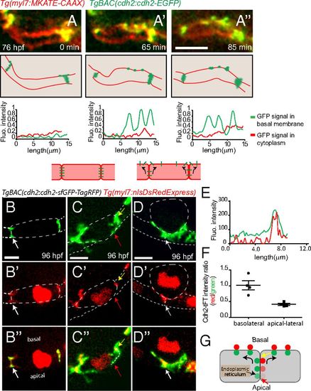
Potential movement of Cdh2-EGFP during cardiac trabeculation. (A-A′′) Spinning disk confocal images of TgBAC(cdh2:cdh2-EGFP);Tg(myl7:MKATE-CAAX) hearts. Cdh2-EGFP appears to relocalize from cell-cell junctions to the basal region of cardiomyocytes, as seen in the single-plane images at different points and depicted in the illustrations here (single-plane images taken from Movie S6). (Bottom) Line scan quantification of Cdh2-EGFP signal in the cytoplasm (red) and basal membrane (green) of cardiomyocytes. (B-D′′) Spinning disk confocal images of TgBAC(cdh2:cdh2-sfGFP-TagRFP);Tg(myl7:nlDsRedExpress) hearts. (B-B′′) Compact layer cardiomyocytes, (C-C′′) delaminating cardiomyocyte, and (D-D′′): delaminated cardiomyocyte; dotted line outlines cardiomyocytes, and the intense red signal in the cytoplasm marks the nucleus. cdh2:Cdh2-tFT expression analysis suggests Cdh2 recruitment to the apical-lateral region of compact layer cardiomyocytes; red arrows in C-C′′ point to an apical-lateral region with newly recruited Cdh2 displaying sfGFP signal without TagRFP signal; white arrows in B-B′′ and D-D′′ point to a region with aged Cdh2 displaying both sfGFP and TagRFP simultaneously. (E) Representative fluorescence intensity distribution of TagRFP and sfGFP in the region depicted by the dotted line in C′′. (F) Scatterplot showing quantification of Cdh2-tFT intensity ratio on the basolateral (yellow arrow in C′′ and G) and apical-lateral (red arrow in C′′ and G) sides of compact layer cardiomyocytes. (G) Diagram depicting hypothetical Cdh2 recruitment to the basal side of the cardiomyocyte plasma membrane from the apical end of cell-cell junctions; the two colors are physically separated for easier visualization. (Scale bars, 5 µm.) |