
Fig. 4
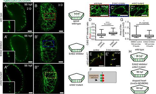
Erbb2 signaling is essential for Cdh2-EGFP localization on the basal side of cardiomyocytes. (A-A′′ and B-B′′) Spinning disk confocal images of TgBAC(cdh2:cdh2-EGFP) hearts. Localization of Cdh2-EGFP in 96 hpf control (wild-type), Erbb2 inhibitor-treated, and erbb2 mutant hearts is shown in the single 2D planes (A-A′′) and 3D reconstructed images (B-B′′). Illustrations of Cdh2-EGFP distribution are shown on the right. (C-C′′) Red, blue, and yellow rectangles shown in the zoomed images are from B-B′′. (D and E) tFT intensity ratio of Cdh2 molecules measured in the regions indicated by the white arrows (E-E′). (F) Schematic diagram depicting Cdh2 localization only at the cell-cell junctions in Erbb2 inhibitor-treated or erbb2 mutant larvae at 96 hpf. (G) Cross-sectional area of Cdh2-EGFP punctae at 96 hpf in DMSO control and Erbb2 inhibitor-treated larvae. (H) Illustration comparing Cdh2 organization in wild-type, stopped, and Erbb2 inhibitor-treated/mutant hearts. (Scale bar: 10 µm in A-A′′, B-B′′, and C-C′′; 5 µm in E and E′.) |