
FIGURE
Fig. S4
Fig. S4
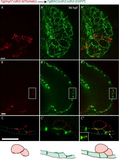
Localization of Cdh2-EGFP during trabeculation. (A-A′′) 3D projection views of Tg(cmlc2:cdh2-tdTomato) donor-derived cardiomyocytes in a TgBAC(cdh2:cdh2-EGFP) host heart. (C-C′′) Zoomed images of the regions outlined by the white box (B-B′′); white arrow points to Cdh2-EGFP between the trabecular and compact cardiomyocytes (C′′). (Scale bars: 10 µm.) |
Expression Data
Expression Detail
Antibody Labeling
Phenotype Data
Phenotype Detail
Acknowledgments
This image is the copyrighted work of the attributed author or publisher, and
ZFIN has permission only to display this image to its users.
Additional permissions should be obtained from the applicable author or publisher of the image.
Full text @ Proc. Natl. Acad. Sci. USA