
Fig. 6
- ID
- ZDB-FIG-160728-6
- Publication
- McCarthy et al., 2016 - An Fgf-Shh signaling hierarchy regulates early specification of the zebrafish skull
- Other Figures
- All Figure Page
- Back to All Figure Page
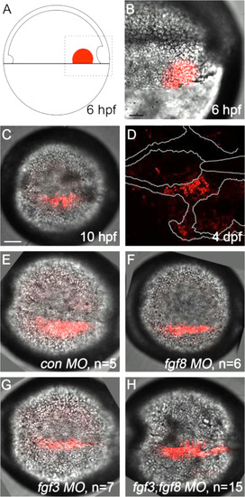
Kaede photoconverted endomesoderm postchordal-progenitor cells migrate appropriately in fgf3;fgf8a knockdown embryos. (A) Schematic of 6 h post-fertilization (hpf) zebrafish embryo showing the region of the embryo photoconverted in all subsequent experiments, dorsal to the right. (B-D) DIC confocal images showing the photoconverted area at 6 hpf (B, magnified region outlined in A), 10 hpf (C), and 4 days post-fertilization (dpf) (D). Cells have migrated adjacent to the notochord by 10 hpf and contributed to the postchordal neurocranium at 4 dpf. At 10 hpf, cells in (E) control, (F) fgf8, (G) fgf3, and (H) fgf3;fgf8 double morpholino-injected embryos are appropriately positioned (compared to C). scale bar=20 µm. |
Reprinted from Developmental Biology, 415(2), McCarthy, N., Sidik, A., Bertrand, J.Y., Eberhart, J.K., An Fgf-Shh signaling hierarchy regulates early specification of the zebrafish skull, 261-77, Copyright (2016) with permission from Elsevier. Full text @ Dev. Biol.