
Fig. S7
- ID
- ZDB-FIG-160728-17
- Publication
- McCarthy et al., 2016 - An Fgf-Shh signaling hierarchy regulates early specification of the zebrafish skull
- Other Figures
- All Figure Page
- Back to All Figure Page
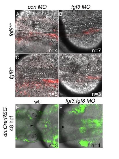
Postchordal-progenitor cells are present adjacent to the notochord at 24 and 48 hpf. (A-D) Confocal images of DIC and Kaede photoconverted postchordal-progenitor cells (in red) at 24 hpf. At 24 hpf, cells in (A) control morpholino- injected wildtype, (B) fgf3 morpholino-injected wildtype , (C) control morpholino-injected fgf8a mutant, and (D) fgf3 morpholino-injected fgf8a mutants are positioned appropriately next to the notochord. Dorsal view with anterior to the left. The notochord is outlined in a white dashed line. (E-F) Confocal and DIC images of tamoxifen-induced drl:Cre;ubi:RSG embryos of (E) wildtype and (F) fgf3;fgf8 MO-injected backgrounds at 48 hpf. Arrowhead denotes anterior notochord. Tamoxifen-induced conversion from red to green shows cells in (F) fgf3;fgf8 MO-injected embryos abutting the notochord at 48 hpf. scale bar= 20 µm. |
Reprinted from Developmental Biology, 415(2), McCarthy, N., Sidik, A., Bertrand, J.Y., Eberhart, J.K., An Fgf-Shh signaling hierarchy regulates early specification of the zebrafish skull, 261-77, Copyright (2016) with permission from Elsevier. Full text @ Dev. Biol.