
Fig. 4
- ID
- ZDB-FIG-160728-4
- Publication
- McCarthy et al., 2016 - An Fgf-Shh signaling hierarchy regulates early specification of the zebrafish skull
- Other Figures
- All Figure Page
- Back to All Figure Page
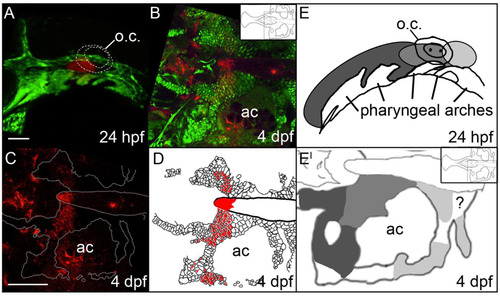
The anterior/posterior organization of postchordal cells is set by 24 h post-fertilization (hpf). (A-C) Confocal images of a Kaede-injected embryo at (A) 24 hpf and (B and C) 4 days post fertilization (dpf). Anterior is to the left. (A) At 24 hpf Kaede was photoconverted, shown in red (the remaining green Kaede is not evident due to the intense green fluorescence from the fli1:EGFP transgene), and (B) at 4 dpf, this same embryo shows labeling in the postchordal neurocranium (inset shows relative position in the neurocranium of magnified view). (C) Red channel only, the neurocrania is outlined in white. (D) Schematic of panel C, with red cells depicting the photoconverted region. (E and E′) Graphical representation of Kaede-mediated fate mapping at 24 hpf and 4 dpf. Inset in E′ shows relative position of magnified neurocrania. Question mark denotes a region that remained unlabeled in our analyses. ac- auditory capsule, o.c.-otic capsule. Scale bars=10 µm in A and 20 µm in C. |
Reprinted from Developmental Biology, 415(2), McCarthy, N., Sidik, A., Bertrand, J.Y., Eberhart, J.K., An Fgf-Shh signaling hierarchy regulates early specification of the zebrafish skull, 261-77, Copyright (2016) with permission from Elsevier. Full text @ Dev. Biol.