|
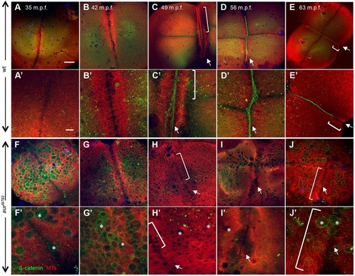
aura mutant embryos do not recruit cell adhesion components to furrows and exhibit aberrant furrow microtubule dynamics. (A-E) In wild-type (12/12), the FMA forms as tubules become arranged in a parallel fashion (A-B′). As the furrow matures, FMA tubules become tilted and accumulate at the furrow distal end (E,E′). During furrow maturation, cell adhesion components such as β-Catenin accumulate at the furrow (C-E). (F-J) In aura mutants, FMA tubules form (F-H, brackets in H,H′) but remain in a parallel array arrangement throughout the length of the furrow (J,J′, brackets), failing to aggregate at the furrow distal ends (assayed at 49-63mpf; 15/15). All aura mutant embryos examined also fail to recruit β-Catenin to the furrow (H-J, arrows). Instead, β-Catenin localizes to ectopic CGs (F′-J′, asterisks). (A′-J′) Higher magnification views of A-J. Scale bars: 100µm in A-J; 10µm in A′-J′.
|