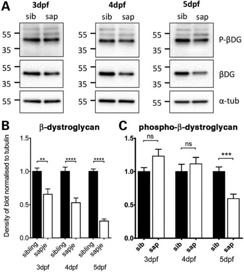
Levels of β-dystroglycan and β-dystroglycan phosphorylated on tyrosine in sapje and sibling larvae. Western blots of lysates of individual 3, 4 and 5 dpf sibling and sapje larvae western blotted with antibodies against phosphorylated β-dystroglycan [p-β-DG, (A) top], non-phosphorylated β-dystroglycan [β-DG, (A) middle] and α-tubulin was used as a loading control [α-tub, (A) bottom]. Numbers represent relative position of molecular weight markers in kDa. (B and C) The integrated density of the blots probed against β-DG and p-β-DG shown in (A), quantified relative to α-tubulin levels in each sample. Graphs show mean + SEM of 12 (B) or 9 (C) samples from three independent experiments in each case. (B) There is a significant decrease in the level of β-dystroglycan in larvae with the sapje mutation at 3, 4 and 5 dpf (unpaired t-tests, 3 dpf: P = 0.0016; 4 dpf: P<0.0001; 5 dpf P < 0.0001). (C) Levels of phosphorylated dystroglycan are slightly increased in sapje at 3 and 4 dpf, but this increase is not statistically significant (P > 0.05). When compared with sibling lysates, levels of phosphorylated dystroglycan at 5 dpf are significantly lower in sapje (P = 0.0007).
|
