|
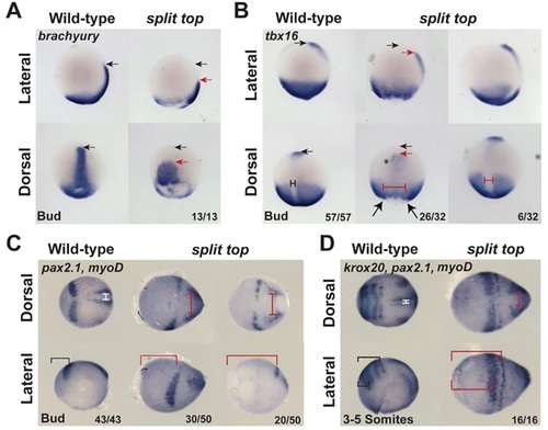
Convergent extension in split top embryos. Expression of (A) brachyury, (B) tbx16, (C,D) pax2.1, (D) krox20 and (C,D) myod indicates an expansion of the midline (probably combined with increased midline mesoderm tissue) and reduced extension in split top mutants. Additionally, whereas krox20 marks rhombomeres 3 and 5 in wild-type embryos, in split top mutants a single krox20 stripe is evident, indicating a loss of either rhombomeres 3 or 5, or a delay in rhombomere 5 expression. Black arrows mark the anterior-most wild-type expression domain and red arrows the anterior-most split top expression domain. The dorsal midline tissue is marked by T-bars in wild-type (black or white) and split top (red) mutants. The brackets indicate the distance from the anterior-most point of the embryo to the neural markers. In A,B, anterior is to top; in C,D, anterior is to left.
|