
Fig. 2
- ID
- ZDB-FIG-160329-12
- Publication
- Chiu et al., 2016 - A Zebrafish Genetic Screen Identifies Neuromedin U as a Regulator of Sleep/Wake States
- Other Figures
- All Figure Page
- Back to All Figure Page
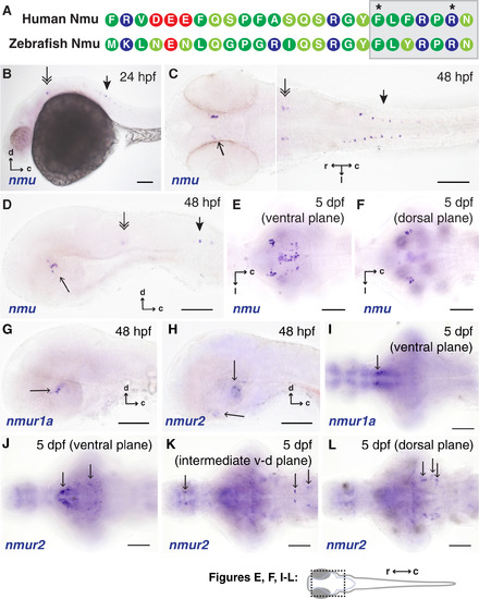
The Sequence of Nmu and Expression of nmu and nmu receptors Are Conserved in Zebrafish (A) The zebrafish predicted Nmu peptide sequence is well conserved with the human Nmu mature peptide. The C-terminal heptapeptide and critical residues are indicated by the shaded box and asterisks. Amino acids are color coded by type (after Malendowicz et al., 2012): blue, basic; red, acidic; green, non-polar; light green, polar. (B-F) Endogenous expression of nmu in the hypothalamus (single arrow), brainstem (double arrow), and spinal cord (arrowhead) of 24-120 hpf zebrafish. (G and H) Lateral views of nmur1a and nmur2 expression at 48 hpf. (I) nmur1a expression in 5 dpf larvae is restricted to the rostro-ventral hypothalamus. (J-L) Widespread but discrete expression of nmur2 in 5 dpf zebrafish brain, starting from a ventral focal plane (J) and ending in a more dorsal focal plane (L). Note that a prominent population of cells in the hypothalamus (left/rostral arrow in J) does not localize to the zebrafish preoptic nucleus, which is not visible in this focal plane. Also note several discrete clusters of cells in the hindbrain (arrows in L), which are located in the vicinity of brainstem arousal systems such as the locus coeruleus. In (G)-(L), arrows distinguish cellular labeling from diffuse background staining. Scale bars = 100 µm. |
| Genes: | |
|---|---|
| Fish: | |
| Anatomical Terms: | |
| Stage Range: | Prim-5 to Day 5 |