
Fig. 2
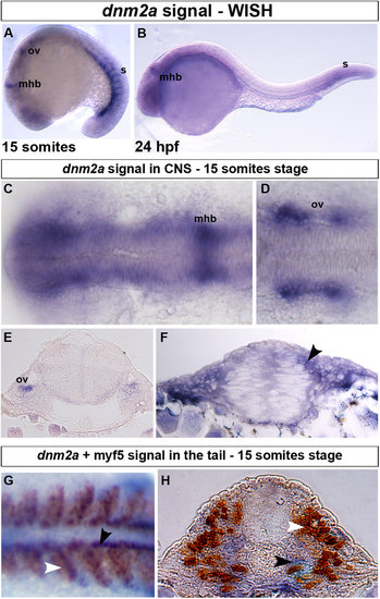
WISH and double-staining of dnm2a and myf5. WISH shows that dnm2a transcripts are present in the CNS and tail of zebrafish embryos from early somitogenesis (11 hpf approximately) to 30 somites stage (24 hpf). (A) At the 15-somite stage dnm2a is present in the tail (somites, s), at the midbrain/hindbrain boundary (mhb) and, bilaterally, at the two otic vesicles (ov). (B) At 24 hpf, dnm2a is present in newly formed somites and appears to be declining in intensity at the midbrain/hindbrain boundary. (C) WISH shows dnm2a signal in CNS, in embryos at 15 somites stage, more pronounced in the midbrain/hindbrain boundary and in the two bilateral otic vesicles (D,E). (F) The expression of dnm2a is diffuse in the neural tube, and more intense in the periventricular and in the dorso-lateral portion (black arrowhead). (G) Double-staining reveals dnm2a and myf5 at the 15 somite stage: dnm2a appears (in dorsal view of flat mounted embryo dnm2a in somites) posteriorly and close to the notochord (black arrowhead) overlapping to some extent with myf5 (white arrowhead); in cross section (H), dnm2a is present in the medio-ventral part of the somite and in several adaxial cells (black arrowhead), while myf5 is expressed in medial cells (white arrowhead). |
| Genes: | |
|---|---|
| Fish: | |
| Anatomical Terms: | |
| Stage Range: | 14-19 somites to Prim-5 |