
FIGURE
Fig. 8
Fig. 8
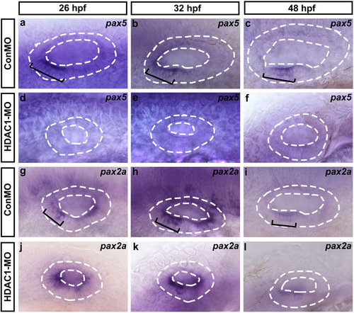
Whole-mount in situ hybridizations to pax5 and pax2a. (a-l) Lateral views of control (ConMO) and HDAC1 morphant otic vesicles at 26 hpf, 32 hpf, and 48 hpf. Black brackets indicate the localization of pax5-expressing (a-c) and pax2a-expressing (g-i) cells as appropriate. Embryos injected with HDAC1 morpholino show reductions in the levels of pax5 (d-f) and pax2a (j-l) expression. The otic vesicles are outlined by dashed lines. All images show lateral views with the anterior to the left and the dorsal side up. |
Expression Data
Expression Detail
Antibody Labeling
Phenotype Data
Phenotype Detail
Acknowledgments
This image is the copyrighted work of the attributed author or publisher, and
ZFIN has permission only to display this image to its users.
Additional permissions should be obtained from the applicable author or publisher of the image.
Full text @ Sci. Rep.