
Fig. 7
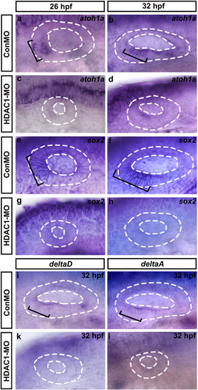
Whole-mount in situ hybridizations to atoh1a, sox2, deltaD, and deltaA. Lateral views of control (ConMO) and HDAC1 morphant otic vesicles at 26 hpf and 32 hpf. Black brackets indicate the regions of atoh1a-expressing (a,b) and sox2-expressing (e,f) cells as appropriate. Embryos injected with HDAC1 morpholino show a reduction in levels of atoh1a (c,d) and sox2 (g,h) expression. (i-l) Whole-mount in situ hybridizations to deltaD and deltaA. Lateral views of control (ConMO) and HDAC1 morphant otic vesicles at 32 hpf. Black brackets indicate the localization of deltaD-expressing (i) and deltaA-expressing (j) cells. Loss of function of HDAC1 reduced the expression of deltaD (k) and deltaA (l). Otic vesicles are outlined by dashed lines. All images show lateral views with the anterior to the left and the dorsal side up. |