
FIGURE
Fig. 3
- ID
- ZDB-FIG-160218-1
- Publication
- Duran et al., 2015 - Collagen duplicate genes of bone and cartilage participate during regeneration of zebrafish fin skeleton
- Other Figures
- All Figure Page
- Back to All Figure Page
Fig. 3
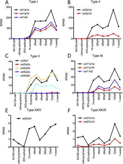
Transcriptional analysis of fibrillar collagens during zebrafish development. Each graph shows expression of all the genes of a collagen type during several developmental stages: 64/128 cells, oblong-sphere, 50% epiboly., 15 somites, 36, 48, 60, and 72 hpf and 1 week post-fertilization. Expression is shown as RPKM. |
Expression Data
| Genes: | |
|---|---|
| Fish: | |
| Anatomical Term: | |
| Stage Range: | 64-cell to Days 7-13 |
Expression Detail
Antibody Labeling
Phenotype Data
Phenotype Detail
Acknowledgments
This image is the copyrighted work of the attributed author or publisher, and
ZFIN has permission only to display this image to its users.
Additional permissions should be obtained from the applicable author or publisher of the image.
Reprinted from Gene expression patterns : GEP, 19(1-2), Duran, I., Csukasi, F., Taylor, S.P., Krakow, D., Becerra, J., Bombarely, A., Marí-Beffa, M., Collagen duplicate genes of bone and cartilage participate during regeneration of zebrafish fin skeleton, 60-9, Copyright (2015) with permission from Elsevier. Full text @ Gene Expr. Patterns