
Fig. 6
- ID
- ZDB-FIG-160212-27
- Publication
- Tomer et al., 2015 - SPED Light Sheet Microscopy: Fast Mapping of Biological System Structure and Function
- Other Figures
- All Figure Page
- Back to All Figure Page
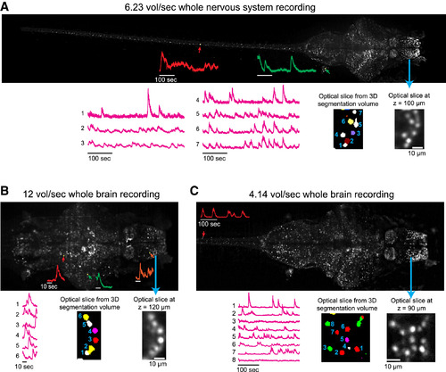
Rapid Cellular-Resolution Functional Mapping of the Entire Larval Zebrafish Nervous System (A–C) The camera-frame-rate limited volumetric imaging speed of SPED light sheet is demonstrated by performing rapid cellular-resolution functional mapping of the nervous system of 10 dpf Tg(elavl3:H2B-GCaMP6s) zebrafish larvae. Three smaller ROIs of the camera frame were used to image: (A) the entire nervous system with a 4×/0.28 NA objective at 6.23 volumes per second (3 mm × 0.5 mm × 0.2 mm, 39 z slices), (B) the whole brain with a 4×/0.28NA objective at 12 volumes per second (0.9 mm × 0.4 mm × 0.2 mm, 40 z slices), and (C) the whole brain and anterior spinal cord with a 10×/0.25NA objective at 4.14 volumes per second (1.2 mm × 0.43 mm × 0.2 mm, 39 z slices). The maximum intensity projection images were generated from a collapsed 3D volume generated by voxel-wise standard deviation (SD) across the entire recording durations. Cellular resolution is demonstrated by several examples of activity traces (ΔF/F versus time) of neurons marked by colored arrows, and of neighboring cells shown in optical slices from respective volumes and their automated 3D segmentation. See Figure S6 for the top 99 example activity traces (ordered according to the variance across time) from the three datasets. Movies S5, S6, S7 exhibit the activity time series (ΔF/F versus time) of these datasets, and Movie S8 shows details of automated 3D segmentation. |
Reprinted from Cell, 163, Tomer, R., Lovett-Barron, M., Kauvar, I., Andalman, A., Burns, V.M., Sankaran, S., Grosenick, L., Broxton, M., Yang, S., Deisseroth, K., SPED Light Sheet Microscopy: Fast Mapping of Biological System Structure and Function, 1796-806, Copyright (2015) with permission from Elsevier. Full text @ Cell