
Fig. 3
- ID
- ZDB-FIG-160205-86
- Publication
- Marsden et al., 2015 - In Vivo Ca(2+) Imaging Reveals that Decreased Dendritic Excitability Drives Startle Habituation
- Other Figures
- All Figure Page
- Back to All Figure Page
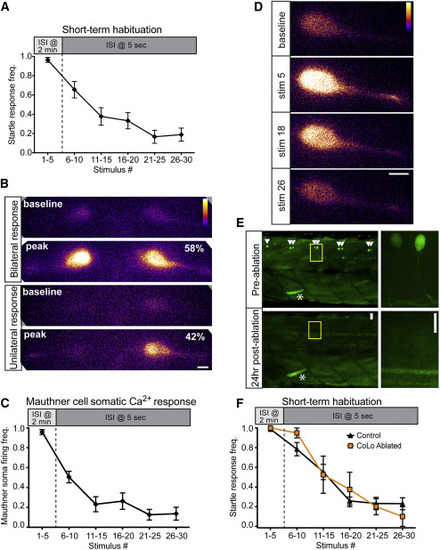
Inhibition Downstream of the M-Cell Does Not Cause Startle Habituation (A) A 30-stimulus assay (5 pulses at 2 min ISI and 25 pulses at 5 s ISI) induced robust startle habituation in head-restrained larvae. Error bars indicate SEM. (B) 40× magnification images of bilateral M-cell GCaMP6s labeling demonstrate unilateral (bottom, 42%) and bilateral (top, 58%) M-cell firing (n = 50 responses from five fish; scale bar, 10 µm). (C) The frequency of M-cell somatic Ca2+ responses during short-term habituation is strongly reduced during habituation. Error bars indicate SEM. (D) Representative images show baseline and peak fluorescence for non-habituating (stim 5) and habituating stimuli (stim 18 and 26; scale bar, 10 µm). (E) Representative images at 10× (left) and 63× (right) show pre-ablation labeling of CoLo interneurons and their absence 24 hr post-ablation. Asterisk highlights a GFP-labeled muscle cell to indicate the same segments are imaged pre- and post-ablation (scale bar 10 µm). (F) Both control and CoLo-ablated larvae rapidly habituate to repeated acoustic stimulation. Error bars indicate SEM. |