
Fig. 11
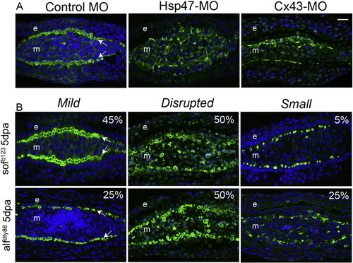
Disruption of actinotrichia following changes in Hsp47 or Cx43 expression. Single plane confocal sections are obtained for transverse sections immunostained with Collagen type II (green) and counterstained for nuclei with To-pro (blue). (A) Fins are injected with standard control MO, Hsp47-MO or Cx43-MO and evaluated at 1 dpe. Col II expression is disrupted following Hsp47 and Cx43 knockdown. (B) Variable actinotrichia phenotypes observed in 5 dpa transverse sections of both sof b123 and alf dty86. The defects were categorized as mild, disrupted organization, or small. Arrows indicate actinotrichia, ‘e’ is epidermis and ‘m’ mesenchyme. Scale bar is 10µm and applies to all panels. |
| Antibody: | |
|---|---|
| Fish: | |
| Condition: | |
| Knockdown Reagents: | |
| Anatomical Term: | |
| Stage: | Adult |
| Fish: | |
|---|---|
| Condition: | |
| Knockdown Reagents: | |
| Observed In: | |
| Stage: | Adult |
Reprinted from Mechanisms of Development, 138 Pt 3, Bhadra, J., Iovine, M.K., Hsp47 mediates Cx43-dependent skeletal growth and patterning in the regenerating fin, 364-74, Copyright (2015) with permission from Elsevier. Full text @ Mech. Dev.