
Fig. 3
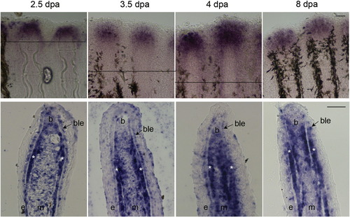
serpinh1b expression over time in regenerating fins. Whole mount in situ hybridization for serpinh1b at different time points on regenerating fins (top). The amputation plane is indicated in 2.5, 3.5 and 4 dpa fins. For 8 dpa fins, the amputation plane is outside the field of view. In situ hybridization on cryosectioned fins at different time points (bottom). Similar localization of serpinh1b gene in basal layer of epithelium (ble) and skeletal precursor cells (*) is observed. Blastema (b), basal layer of epidermis (ble), and skeletal precursor cells (*). The scale bars are 50µm. The scale bar in the upper 8 dpa panel applies to all panels for whole mount images. The scale bar in the lower 8 dpa panel applies to all panels for cryosections. |
| Gene: | |
|---|---|
| Fish: | |
| Condition: | |
| Anatomical Terms: | |
| Stage: | Adult |
Reprinted from Mechanisms of Development, 138 Pt 3, Bhadra, J., Iovine, M.K., Hsp47 mediates Cx43-dependent skeletal growth and patterning in the regenerating fin, 364-74, Copyright (2015) with permission from Elsevier. Full text @ Mech. Dev.