
Fig. S4
- ID
- ZDB-FIG-151214-74
- Publication
- Askary et al., 2015 - Iroquois Proteins Promote Skeletal Joint Formation by Maintaining Chondrocytes in an Immature State
- Other Figures
- All Figure Page
- Back to All Figure Page
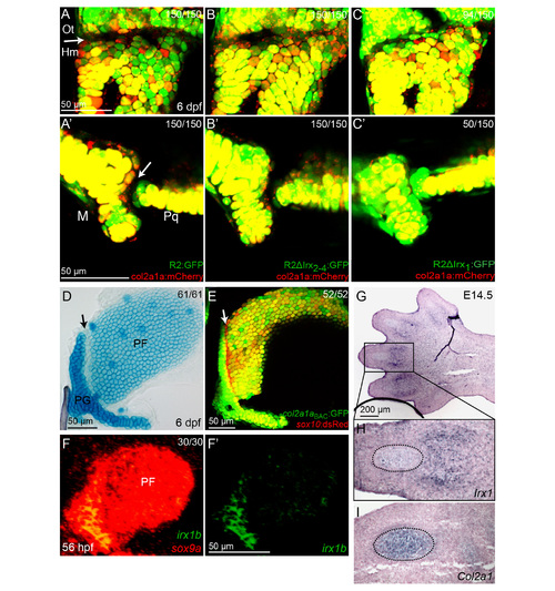
Potential roles of Irx genes at multiple joints. (A-C) Modified R2 enhancer transgenes (green) are shown relative to a wild-type col2a1aBAC:mCherry-NTR transgene (red) which labels chondrocytes but is excluded from joints (arrows). Compared to the control wild type R2 (A,A′), expression from an R2 enhancer lacking the first Irx site is expanded into the hyomandibula – (Hm) otic capsule (Ot) joint (C) and jaw joint (C′). In contrast, loss of the second through fourth Irx sites has no effect on R2 enhancer expression at these joints (B,B′). Experimental numbers are pooled for three independent stable transgenic lines. 50 embryos were examined for each line. M, Meckel’s lower jaw cartilage; Pq, palatoquadrate upper jaw cartilage. (D-F) The joint between the pectoral fin (PF) and pectoral girdle (PG) shares some of the features of the hyoid joint. At 6 dpf, cells within the pectoral fin joint stain poorly with Alcian Blue relative to surrounding chondrocytes (D, arrow). The pectoral fin joint (arrow) also expresses much lower levels of col2a1aBAC:GFP relative to surrounding chondrocytes, while sox10:dsRed (red) uniformly labels chondrocytes (E). At 56 hpf, irx1b is expressed within precursors of the pectoral fin joint (F). sox9a expression (red) labels early chondrocytes at this stage. Numbers indicate proportion of animals showing the displayed phenotype. (G-I) in situ hybridization shows expression of Irx1 in the digit joints in an E14.5 mouse hindpaw. Magnifications of adjacent sections (H,I) show that Irx1 and Col2a1 are expressed in a largely mutually exclusive pattern, with Col2a1 but not Irx1 marking cartilage condensations (dashed lines). Serial sections of 2 animals gave similar results. |
Reprinted from Developmental Cell, 35, Askary, A., Mork, L., Paul, S., He, X., Izuhara, A.K., Gopalakrishnan, S., Ichida, J.K., McMahon, A.P., Dabizljevic, S., Dale, R., Mariani, F.V., Crump, J.G., Iroquois Proteins Promote Skeletal Joint Formation by Maintaining Chondrocytes in an Immature State, 358-65, Copyright (2015) with permission from Elsevier. Full text @ Dev. Cell