
Fig. 4
- ID
- ZDB-FIG-151214-71
- Publication
- Askary et al., 2015 - Iroquois Proteins Promote Skeletal Joint Formation by Maintaining Chondrocytes in an Immature State
- Other Figures
- All Figure Page
- Back to All Figure Page
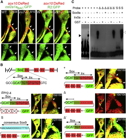
Irx Genes Directly Repress a col2a1a Enhancer (A) Relative to sox10:dsRed+ chondrocytes (red), col2a1aBAC:GFP, and R2:GFP transgenes (green) show increased col2a1a-driven expression in hyoid joint cells (arrows) of irx7el538 mutants compared with WT siblings. (B) Schematics show predicted Sox and Irx binding sites within the R2 enhancer of the col2a1a gene, as well as modified transgenic constructs. Confocal images at 6 dpf show the connection of the hyosymplectic and ceratohyal cartilages, with magnified images showing the bipartite hyoid joints (arrows). In all panels, the WT col2a1aBAC:mCherry-NTR transgene (red) displays lower levels in hyoid joints cells than neighboring chondrocytes. Transgenes with WT or modified R2 enhancers driving GFP are shown in green. Both the WT R2 enhancer and a version missing the second through fourth putative Irx sites are similar to WT col2a1aBAC:mCherry-NTR in being excluded from joints (yellow reflects overlap). Loss of the overlapping Sox/Irx site of the R2 enhancer abolishes most cartilage expression (red), and replacement with a Sox9 consensus site or deletion of just the Irx half-site results in ectopic joint expression (green). In the case of R2 enhancer missing the overlapping Sox/Irx site (Δ) 104 animals injected with the plasmid show similar expression and out of 30 founders screened, none showed more than just a sparse GFP expression in a few cartilage cells. For other panels experimental numbers are pooled for three independent stable transgenic lines. 50 embryos were examined for each line. Numbers indicate the proportion of animals showing the displayed phenotype. Scale bars represent 30 µM. (C) An electrophoretic mobility shift assay shows binding of Irx5a (closed arrowhead) and Sox9a (open arrowhead) but not a control GST protein to a probe containing the wild-type Sox/Irx site (+). Sox9a binds a probe deleted for the Irx half-site (Δ′) but not the entire Sox/Irx site (Δ), and binding is restored by replacement of the Sox/Irx site with a consensus Sox9 binding site (S). See also Figure S4. |
| Genes: | |
|---|---|
| Fish: | |
| Anatomical Terms: | |
| Stage: | Day 6 |
| Fish: | |
|---|---|
| Observed In: | |
| Stage: | Day 6 |
Reprinted from Developmental Cell, 35, Askary, A., Mork, L., Paul, S., He, X., Izuhara, A.K., Gopalakrishnan, S., Ichida, J.K., McMahon, A.P., Dabizljevic, S., Dale, R., Mariani, F.V., Crump, J.G., Iroquois Proteins Promote Skeletal Joint Formation by Maintaining Chondrocytes in an Immature State, 358-65, Copyright (2015) with permission from Elsevier. Full text @ Dev. Cell