
Fig. S1
- ID
- ZDB-FIG-151214-72
- Publication
- Askary et al., 2015 - Iroquois Proteins Promote Skeletal Joint Formation by Maintaining Chondrocytes in an Immature State
- Other Figures
- All Figure Page
- Back to All Figure Page
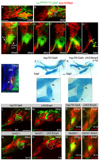
Progressive restriction of irx7:GFP to the hyoid joint and Hand2-independent inhibition of irx7 expression by Bmp signaling. (A-H) Confocal projections of the mandibular and hyoid arches at 24 (A), 38 (B), and 48 hpf (C) and the hyoid cartilages at 56 hpf onwards (D-H). sox10:mCherryCAAX (24, 48, 56, and 80 hpf and 9 dpf) and sox10:dsRed (38, 60, and 68 hpf), collectively abbreviated as “sox10:Red”, mark neural-crest-derived arch mesenchyme and later chondrocytes in red. The SAGp11A line is an insertion of a GFP-containing retrotransposon into the second exon of the irx7 gene. irx7-driven GFP expression is seen weakly throughout the hyoid arch at 24 hpf (A) and then becomes refined to the intermediate domain of the hyoid arch by 38 hpf (B). Expression then increases in the hyoid joint and adjacent symplectic cartilage region by 48 hpf (C). At later times, GFP expression persists around the hyoid joint but is gradually lost in the growing symplectic cartilage (D-G). In mid-stage larvae (9 dpf), GFP is highly restricted to chondrocytes at either end of the bipartite hyoid joint (arrows), as well as in perichondral cells (arrowhead) lining the joint (H). (I) At 72 hpf, relative to sox10:GFPCAAX+ cartilages (anti-GFP, blue), overlapping expression of irx7 (red) and irx5a (green) is seen at the developing hyoid joint (arrow). irx5a is also expressed in posterior hyoid arch cells (asterisk). (J,K) Alcian blue labeling of cartilage in control hsp70I:Gal4 and hsp70I:Gal4; UAS:Bmp4 animals subjected to a heat shock from 40-44 hpf. Misexpression of Bmp4 at this stage causes joint fusions (arrows, see magnification of hyoid joint) in the absence of the large-scale dorsoventral patterning defects caused by earlier (20-24 hpf) induction of Bmp4 (Zuniga et al., 2011). (L-O) Control hsp70I:Gal4 (L), hsp70I:Gal4; UAS:Bmp4 (M), hand2-/- (N), and hand2-/-; hsp70I:Gal4; UAS:Bmp4 (O) embryos subjected to a heat shock from 20-24 hpf. In situ hybridization shows ventral expansion (brackets) of irx7 expression in hand2-/- embryos, and loss of hyoid joint expression (dashed polygons) upon Bmp4 misexpression in both wild-type and hand2 mutant backgrounds. As a reference, dlx2a expression labels mandibular and hyoid arch mesenchyme in red. (P-S) Control hsp70I:Gal4 (P), hsp70I:Gal4; UAS:Bmp4 (Q), hand2-/- (R), and hand2-/-; hsp70I:Gal4; UAS:Bmp4 (S) embryos subjected to a heat shock from 40-44 hpf. In situ hybridization shows ventral expansion of irx7 expression in hand2-/- embryos, and loss of hyoid joint expression upon Bmp4 misexpression. Loss of hand2 partially restores some irx7 expression upon Bmp4 misexpression, suggesting that Bmp4 may act through both Hand2 and other effectors at this later stage. As a reference, sox9a expression labels newly forming chondrocytes in red. D, dorsal; I, intermediate; V, ventral; Hm, hyomandibula. Numbers indicate proportion of animals showing the displayed phenotype. Scale bars = 50 µM. |
Reprinted from Developmental Cell, 35, Askary, A., Mork, L., Paul, S., He, X., Izuhara, A.K., Gopalakrishnan, S., Ichida, J.K., McMahon, A.P., Dabizljevic, S., Dale, R., Mariani, F.V., Crump, J.G., Iroquois Proteins Promote Skeletal Joint Formation by Maintaining Chondrocytes in an Immature State, 358-65, Copyright (2015) with permission from Elsevier. Full text @ Dev. Cell