
Fig. S1
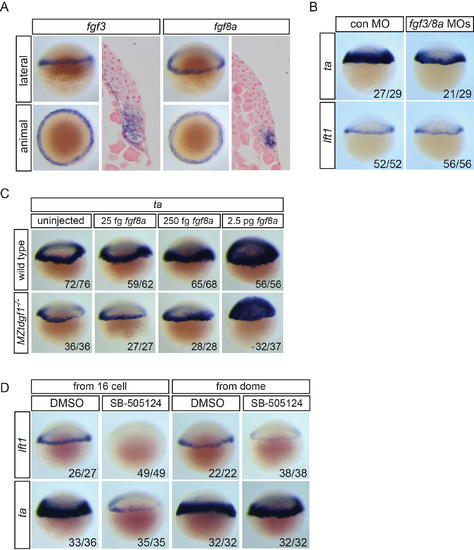
Fgf signaling inhibition and Nodal signaling (A) Expression of fgf3 and fgf8a in 40% epiboly embryos in the margin shown as WISH and sections. (B) ta and lft1 expression in 40% epiboly embryos injected with control MO or a combination of fgf3 and fgf8a MOs. (C) ta expression in wild type and maternal-zygotic (MZ) tdgf1-/- mutants. fgf8a mRNA (25 fg, 250 fg or 2.5 pg) was injected into wild type or MZtdgf1-/- mutant embryos, which were assayed by WISH for ta expression at 40% epiboly. ta is induced equally well in both backgrounds with 2.5 pg fgf8a mRNA. (D) lft1 and ta expression following inhibition of Nodal signaling with SB-505124 from the 16-cell or dome stage. When inhibited from dome stage, lft1 expression is severely reduced, whereas ta expression is unaffected. |
Reprinted from Developmental Cell, 35, van Boxtel, A.L., Chesebro, J.E., Heliot, C., Ramel, M.C., Stone, R.K., Hill, C.S., A Temporal Window for Signal Activation Dictates the Dimensions of a Nodal Signaling Domain, 175-185, Copyright (2015) with permission from Elsevier. Full text @ Dev. Cell