
Fig. S2
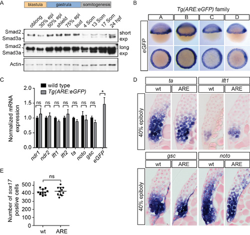
Generation of the Tg(ARE:eGFP) transgenic line (A) Western blot for Smad2 and Smad3a/b during development. Smad3a/b are not expressed at appreciable levels until somitogenesis stages, whereas Smad2 is expressed at all stages shown. A long and short exposure (exp) of the same blot is shown. Actin is a loading control. (B) WISH for eGFP in four Tg(ARE:eGFP) lines obtained from independent founders (labeled A–D). Note that individual lines differ slightly in strength of staining and background. (C) qPCR on pooled 40% epiboly wild type and Tg(ARE:eGFP) embryos for the indicated genes normalized to eef1a1l1. Means ± SEM are shown (* t-test: P < 0.05, n=5). ns, not significant. No significant differences were found for any of the genes tested, except eGFP. (D) Sections of WISH-stained 40% epiboly embryos. No differences were found in the extent of ta, lft1, gsc or noto (flh) staining between wild type and Tg(ARE:eGFP) embryos. Note that lateral sections are shown for ta and lft1, whilst dorsal sections are shown for gsc and noto. (E) Comparison of the number of sox17 positive cells in 75% epiboly wild type and Tg(ARE:eGFP) embryos. No significant difference was found between the number of endodermal cells between the two lines using a Mann-Whitney U test. In (D) and (E) wt, wild type; ARE, Tg(ARE:eGFP). |
Reprinted from Developmental Cell, 35, van Boxtel, A.L., Chesebro, J.E., Heliot, C., Ramel, M.C., Stone, R.K., Hill, C.S., A Temporal Window for Signal Activation Dictates the Dimensions of a Nodal Signaling Domain, 175-185, Copyright (2015) with permission from Elsevier. Full text @ Dev. Cell