
Fig. 4
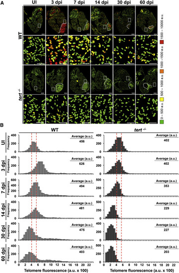
Heart Cryoinjury Induces a Telomerase-Dependent Increase in Telomere Reserves (A) Representative telomap images of WT and tert-/- heart, uninjured (UI) or fixed at different days postinjury [dpi]. Nuclei are assigned a four-color code according to their average telomere fluorescence in a.u. The cells with the longest telomeres are visualized in red, and the cells with the shortest telomeres are presented in green. Dotted lines mark the injured area. The second and fourth rows show higher magnifications of the boxed areas highlighted in the entire heart section images. Scale bars, 100 µm and 10 µm for magnification images. (B) Telomere fluorescence frequency histograms of cardiac WT and tert-/- cells in uninjured hearts and hearts at 3, 7, 14, 30, and 60 days after cryoinjury. Mean telomere fluorescence is indicated in a.u. The injury site was excluded from the analysis. Note the increase in the number of cells with long telomeres at 3 dpi in WT hearts, followed by reestablishment of the initial profile. This response is lacking in tert/ cells, and, at later stages, cells have shorter telomeres than initially. See also Figure S6. |