
Fig. S3
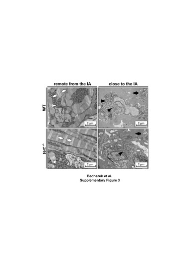
Cardiomyocyte dedifferentiation is unaffected in the absence of telomerase. Transmission electron micrographs of cardiomyocytes at 3 days post injury. (Left) Representative cardiomyocytes away from the injury site in WT and tert-/- animals, showing ordered sarcomeric fibers with visible Z-lines (white arrows) and perinuclear mitochondria with typical structure (white arrowheads). (Right) Representative cardiomyocytes close to the injury site, showing disorganized sarcomeric structure with loss of Z-lines (black arrows) and dysmorphic mitochondria (black arrowheads). IA, injury area; WT, wildtype. Scale bars: 2 µm. Related to Figure 2. |