
Fig. 2
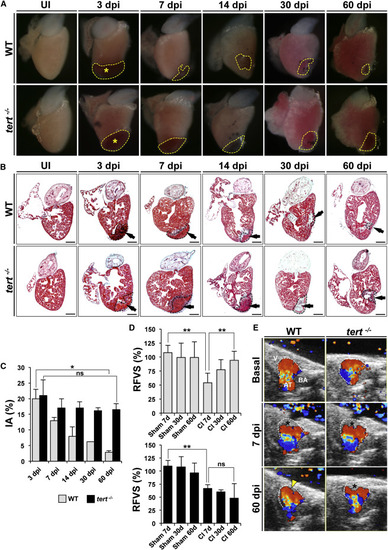
Heart Regeneration Is Inhibited Strongly in tert-/- Animals (A) Whole-mount views of uninjured and cryoinjured WT and tert-/- zebrafish hearts dissected at the indicated times post-injury. Dotted lines outline the injured area. Asterisks mark the initial injury site. (B) Masson-Goldner trichrome staining of sagittal sections of uninjured WT and tert-/- hearts at the indicated days after cryoinjury. Dotted black lines outline the injured area. Arrows mark the initial injury site. Scale bars, 100 µm. (C) Size of the ventricle injury (injured area [IA]) on sagittal heart sections at the indicated times post-injury, presented as the percentage of the total ventricular area. Data are means of at least 4 sections/heart from 3-8 hearts/time point, with the exception of WT 30 dpi, where only one heart was analyzed. Data are means ± SEM. p < 0.05 (Mann-Whitney test). (D) Relative FVS (RFVS) in WT and tert/ zebrafish hearts under the basal condition (sham-operated animals) and at the indicated times post-injury. WT hearts recover ventricular function over time but tert-/- hearts do not. Data are means ± SEM of a pool of 10 animals/condition. p < 0.01 (one-way ANOVA followed by Tukey’s honest significant difference test). (E) Representative Doppler echocardiography images of intracardial blood flow in WT and tert-/- hearts in the absence of injury and at the indicated times post-injury. Asterisks mark the initial injury site. See also Movies S1, S2, S3, S4, S5, and S6. See also Figures S1–S3, and Table S1. |