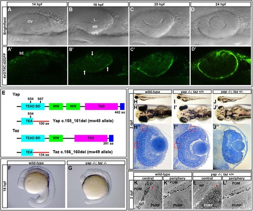
A Yap/Taz-Tead reporter transgene is dynamically expressed during optic cup morphogenesis and yap-/- mutants exhibit RPE defects. (A-D′) Images of live zebrafish from 14-24hpf showing optic cup development and 4xGTIIC:d2GFP transgene expression (green). Arrows indicate cells that are expressing the transgene while undergoing morphogenesis. (E) Schematic of wild-type and mutant Yap and Taz. Yap S54 is an essential residue for Tead binding and S87 is phosphorylated by Lats leading to cytoplasmic retention. The Yap c.158_161del and Taz c.156_160del mutants contain frameshifts resulting in early stop codons. TEAD BD, Tead transcription factor binding domain (light blue); WW, dual tryptophan motif (green); TAD, transactivation domain (fushia); PDZ domain (dark blue). (F,G) yap-/-; taz-/- embryos arrest by 18hpf with multiple defects. (H-J′′) Live embryos (H-J′) and sections (H′′,I′′,J′′) of yap-/-; taz+/+ (I-I′′) and yap-/-; taz+/- (J-J′′) showing RPE defects and additional NR defects in yap-/-; taz+/- mutants (J′) compared with control (H-H′′). Boxed areas indicate locations of TEM analysis. (K-L′) Transmission electron microscopy analysis showing areas of normal RPE development (L′) and areas devoid of RPE (L) in yap-/- eyes. Asterisk indicates the presence of primary cilia on neuroepithelial cells. L, lens; OV, optic vesicle; NR, neural retina; RPE, retinal pigment epithelium; SE, surface ectoderm; POM, periocular mesenchyme; NP, neuropil; PhRP, photoreceptor progenitors.
|
