
Fig. 3
- ID
- ZDB-FIG-150831-9
- Publication
- Iyengar et al., 2015 - Poised Regeneration of Zebrafish Melanocytes Involves Direct Differentiation and Concurrent Replenishment of Tissue-Resident Progenitor Cells
- Other Figures
- All Figure Page
- Back to All Figure Page
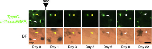
Unpigmented mitfa-Expressing Cells Give Rise to Differentiated Melanocytes by Direct Differentiation during Regeneration Lineage tracing of unpigmented mitfa-expressing cells on the flank of an adult Tg(miniCoopR-mitfa:nlsEGFP) zebrafish before and after neocuproine-mediated ablation of differentiated melanocytes. Unpigmented mitfa-expressing cells (yellow arrowheads) directly differentiated into pigmented melanocytes (white arrowheads). Differentiation was noted by the onset of melanization. Fish were treated with epinephrine only on day 0 prior to imaging. mC, miniCoopR; BF, brightfield; neo, neocuproine. Scale bar, 50 µM. See also Figure S3. |
Reprinted from Developmental Cell, 33(6), Iyengar, S., Kasheta, M., Ceol, C.J., Poised Regeneration of Zebrafish Melanocytes Involves Direct Differentiation and Concurrent Replenishment of Tissue-Resident Progenitor Cells, 631-43, Copyright (2015) with permission from Elsevier. Full text @ Dev. Cell