
Fig. 6
- ID
- ZDB-FIG-150831-12
- Publication
- Iyengar et al., 2015 - Poised Regeneration of Zebrafish Melanocytes Involves Direct Differentiation and Concurrent Replenishment of Tissue-Resident Progenitor Cells
- Other Figures
- All Figure Page
- Back to All Figure Page
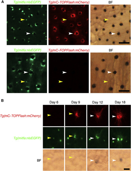
Wnt Signaling Is Activated during Melanocyte Regeneration (A) Flank of an adult zebrafish coexpressing mitfa:nlsEGFP and miniCoopR-TOPFlash:mCherry (top) or mitfa:nlsEGFP and miniCoopR-FOPFlash:mCherry (bottom). In animals with Tg(miniCoopR-TOPFlash:mCherry), mCherry signal was evident in differentiated melanocytes (white arrowheads) but not in unpigmented mitfa-expressing cells (yellow arrowheads). No mCherry signal was observed in Tg(miniCoopR-FOPFlash:mCherry) animals. Fish were treated with epinephrine prior to imaging. mC, miniCoopR; BF, brightfield. Scale bars, 100 µM. (B) Lineage tracing of unpigmented mitfa-expressing cells on the flank of an adult zebrafish coexpressing mitfa:nlsEGFP and miniCoopR-TOPFlash:mCherry after neocuproine treatment. An unpigmented mitfa-expressing cell (yellow arrowheads) beginning to express mCherry shortly before differentiation (white arrowheads). mC, miniCoopR; BF, brightfield. Scale bar, 50 µM. See also Figure S5. |
Reprinted from Developmental Cell, 33(6), Iyengar, S., Kasheta, M., Ceol, C.J., Poised Regeneration of Zebrafish Melanocytes Involves Direct Differentiation and Concurrent Replenishment of Tissue-Resident Progenitor Cells, 631-43, Copyright (2015) with permission from Elsevier. Full text @ Dev. Cell