
Fig. 3
- ID
- ZDB-IMAGE-150831-3
- Publication
- Iyengar et al., 2015 - Poised Regeneration of Zebrafish Melanocytes Involves Direct Differentiation and Concurrent Replenishment of Tissue-Resident Progenitor Cells
- All Figures
- Figures for Iyengar et al., 2015
|
Fig. 3
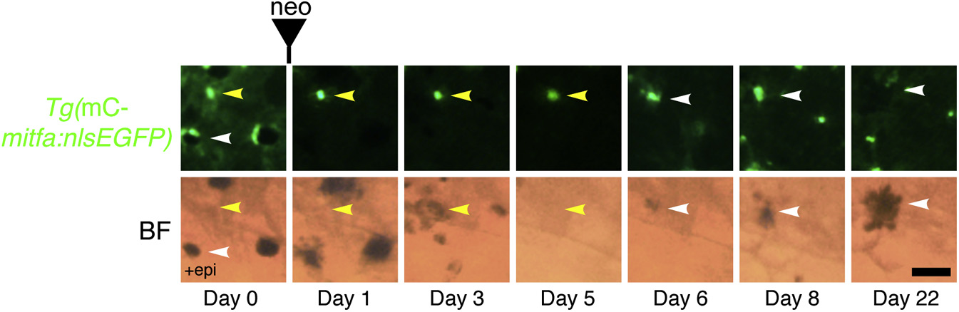
Unpigmented mitfa-Expressing Cells Give Rise to Differentiated Melanocytes by Direct Differentiation during Regeneration
Lineage tracing of unpigmented mitfa-expressing cells on the flank of an adult Tg(miniCoopR-mitfa:nlsEGFP) zebrafish before and after neocuproine-mediated ablation of differentiated melanocytes. Unpigmented mitfa-expressing cells (yellow arrowheads) directly differentiated into pigmented melanocytes (white arrowheads). Differentiation was noted by the onset of melanization. Fish were treated with epinephrine only on day 0 prior to imaging. mC, miniCoopR; BF, brightfield; neo, neocuproine. Scale bar, 50 µM. See also Figure S3.
Reprinted from Developmental Cell, 33(6), Iyengar, S., Kasheta, M., Ceol, C.J., Poised Regeneration of Zebrafish Melanocytes Involves Direct Differentiation and Concurrent Replenishment of Tissue-Resident Progenitor Cells, 631-43, Copyright (2015) with permission from Elsevier. Full text @ Dev. Cell