
Fig. 2
- ID
- ZDB-FIG-150831-8
- Publication
- Iyengar et al., 2015 - Poised Regeneration of Zebrafish Melanocytes Involves Direct Differentiation and Concurrent Replenishment of Tissue-Resident Progenitor Cells
- Other Figures
- All Figure Page
- Back to All Figure Page
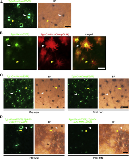
Unpigmented mitfa-Expressing Cells Are Present at the Onset of Regeneration (A) Flank of adult zebrafish expressing miniCoopR-mitfa:nlsEGFP. Unpigmented nlsEGFP-positive nuclei (yellow arrowheads) were admixed with nuclei of differentiated melanocytes (white arrowheads). mC, miniCoopR; BF, brightfield. Scale bar, 100 µM. (B) Flank of adult zebrafish coexpressing mitfa:nlsEGFP and miniCoopR-mitfa:mCherryCAAX. Unpigmented cells with nlsEGFP-positive nuclei (yellow arrowheads) were distinct from differentiated melanocytes (white arrowheads). mC, miniCoopR. Scale bar, 100 µM. (C) Flank of adult zebrafish expressing miniCoopR-mitfa:nlsEGFP before (left) and after (right) neocuproine treatment. Unpigmented cells with nlsEGFP-positive nuclei (yellow arrowheads) persisted whereas differentiated melanocytes (white arrowheads) died after neocuproine treatment. n = 5 fish; representative images are shown. mC, miniCoopR; BF, brightfield; neo, neocuproine. Scale bar, 100 µM. (D) Flank of adult p53(lf) zebrafish coexpressing miniCoopR-mitfa:NTR and mitfa:nlsEGFP before (left) and after (right) Mtz treatment. Unpigmented cells with nlsEGFP-positive nuclei (yellow arrowheads) were spared while differentiated melanocytes (white arrowheads) were ablated following Mtz treatment. n = 3 fish; representative images are shown. Fish were treated with epinephrine prior to imaging. mC, miniCoopR; BF, brightfield. Scale bar, 100 µM. See also Figure S2. |
Reprinted from Developmental Cell, 33(6), Iyengar, S., Kasheta, M., Ceol, C.J., Poised Regeneration of Zebrafish Melanocytes Involves Direct Differentiation and Concurrent Replenishment of Tissue-Resident Progenitor Cells, 631-43, Copyright (2015) with permission from Elsevier. Full text @ Dev. Cell