
FIGURE
Fig. 10
- ID
- ZDB-FIG-150504-35
- Publication
- Quan et al., 2015 - Comparative Distribution and In Vitro Activities of the Urotensin II-Related Peptides URP1 and URP2 in Zebrafish: Evidence for Their Colocalization in Spinal Cerebrospinal Fluid-Contacting Neurons
- Other Figures
- All Figure Page
- Back to All Figure Page
Fig. 10
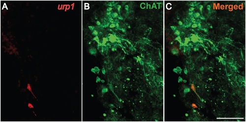
urp1+ cells in the hindbrain are cholinergic neurons expressing ChAT in adult zebrafish. urp1 expression revealed by fluorescent ISH (TAMRA, red) on coronal sections of adult brain, together with a fluorescent immunostaining for ChAT (Alexa 488, green). urp1+ cells express ChAT. Scale bars: 100 µm. |
Expression Data
| Genes: | |
|---|---|
| Fish: | |
| Anatomical Term: | |
| Stage: | Adult |
Expression Detail
Antibody Labeling
Phenotype Data
Phenotype Detail
Acknowledgments
This image is the copyrighted work of the attributed author or publisher, and
ZFIN has permission only to display this image to its users.
Additional permissions should be obtained from the applicable author or publisher of the image.
Full text @ PLoS One