
Fig. 7
- ID
- ZDB-FIG-150504-32
- Publication
- Quan et al., 2015 - Comparative Distribution and In Vitro Activities of the Urotensin II-Related Peptides URP1 and URP2 in Zebrafish: Evidence for Their Colocalization in Spinal Cerebrospinal Fluid-Contacting Neurons
- Other Figures
- All Figure Page
- Back to All Figure Page
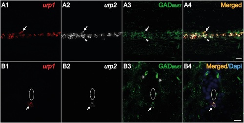
Both urp1+ and urp2+ cells in the spinal cord are GABAergic neurons in adult zebrafish. Simultaneous expression of urp1 and urp2 revealed by double fluorescent ISH (TAMRA, red for urp1 and Cy5, white for urp2) in adult spinal cord, coupled to a fluorescent immunostaining for GAD65/67 (Alexa Fluor 488, green) (A, B). Both urp1+ and urp2+ cells are GAD+ (A, B). Arrows designate triple-stained cells (A, B). Note the occurrence of some doubly-positive cells (urp2/GAD) that do not contain any urp1 (arrowhead) (A). Asterisks designate GABAergic interneurons located at the dorsal part of the spinal cord. The white dash line indicates the central canal. A, sagittal section with dorsal up; B, coronal section with dorsal up. Scale bars: 15 µm. |
| Genes: | |
|---|---|
| Antibody: | |
| Fish: | |
| Anatomical Term: | |
| Stage: | Adult |