
Fig. 8
- ID
- ZDB-FIG-150504-33
- Publication
- Quan et al., 2015 - Comparative Distribution and In Vitro Activities of the Urotensin II-Related Peptides URP1 and URP2 in Zebrafish: Evidence for Their Colocalization in Spinal Cerebrospinal Fluid-Contacting Neurons
- Other Figures
- All Figure Page
- Back to All Figure Page
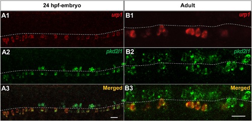
urp1+ cells express pkd2l1, a specific marker of spinal cerebrospinal fluid- contacting neurons in zebrafish. Simultaneous expression of urp1 and pkd2l1 revealed by double fluorescent ISH (TAMRA, red for urp1 and FITC, green for pkd2l1) on 24 hpf-embryo (A) and adult spinal cord sections (B). pkd2l1 mRNA is distributed in two rows of cells along the rostro-caudal axis of the spinal cord both in embryo and adult (A2, B2). All the urp1+ cells are pkd2l1+ (A1,3, B1,3) but only the ventral pkd2l1+ cells are urp1+. The white dash line indicates the central canal. A, lateral views; B, sagittal sections with dorsal up. Scale bars: 20µm. |
| Genes: | |
|---|---|
| Fish: | |
| Anatomical Terms: | |
| Stage Range: | Prim-5 to Adult |