
Fig. 7
- ID
- ZDB-FIG-150430-15
- Publication
- Luz et al., 2014 - Dynamic Association with Donor Cell Filopodia and Lipid-Modification Are Essential Features of Wnt8a during Patterning of the Zebrafish Neuroectoderm
- Other Figures
- All Figure Page
- Back to All Figure Page
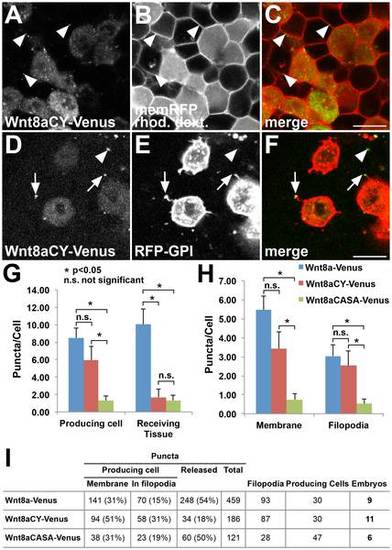
Non-functional Wnt8a mutations have distinct effects on Wnt8a distribution. (A–C) Wnt8aCY-Venus expressing cells colabeled with rhodamine dextran transplanted into a host embryo expressing memRFP. (A) Wnt8aCY-Venus in the expressing cells produce less punctate structures (compare to Fig.1A–C) which nevertheless colocalize with the plasma membrane of receiving cells (arrowheads). (D–F) Mosaic coexpression of Wnt8aCY-Venus and RFP-GPI. The majority of Wnt8aCY-Venus puncta colocalize with filopodia of producing cells (arrow). Only few Wnt8aCY-Venus puncta in the receiving tissue are observed (arrowheads). (G–H) Quantification of the number of puncta in producing cells and in receiving tissue, number of puncta in membrane and in filopodia for Wnt8a-Venus, Wnt8aCY-Venus and Wnt8aCASA-Venus (n = 9 embryos for Wn8-Venus, n = 11 embryos for Wn8-VenusCY, n = 6 embryos for Wnt8aCASA-Venus, error bars represent s.e.m., *p<0.05, n.s. not significant). The number of puncta was normalized to the number of producing cells (labeled with RFG-GPI) in each image. (I) Total numbers for the different parameters analyzed in the quantifications. Producing cells number refers to the sum of RFP-GPI labeled cells in the total number of embryos. (A,D) Green channel, (B,E) red channel, (C,F) overlay. Scale bars: 20 µm. Single confocal planes are shown. |